| SINAE carries out research and developmental projects with public and prívate funds, though the collaboration with public entities such as Pablo Olavide University and Andalusian Center of Developmental Biology (CSIC), and private national and international entities such as Jones Institute, in the Eastern Virginia Medical School (Norfolk, Virginia, USA), OriginElle (Montreal, Canada), Cegyr (Buenos Aires, Argentina), Reprogenetics (Livinstone, New Jersey, USA), Recombine EU, (Bilbao), IVF Spain (Alicante), Ginemed (Seville) and Miguel Servet Hospital (Zaragoza). The development of a new diagnostic method for endometrial receptivityEndometrium is a tissue that suffers big periodic changes along menstrual cycle, and these changes are guided by steroid hormones regulation and paracrine molecules secreted from neighbouring cells. The cycle of endometrium can be divided in three phases: proliferative phase, which correspond to ovarian follicular phase, secretory phase, corresponding to ovarian luteal phase, and menstrual phase. In order to achieve success in embryonic implantation, it must be a synchrony between embryonic development and the endometrium. The endometrial competence to embryonic implantation in blastocyst stage is defined as “receptive”, and that occurs during few days in each menstrual cycle, induced by the increase of oestrogen and progesterone concentrations in medium secretory phase. This period of time, optimum for maternal-embryonic interaction, is known as window of implantation (WOI). It occurs on days 20 and 21 of menstrual cycle, seven days after endogenous LH peak that triggers ovulation. | |
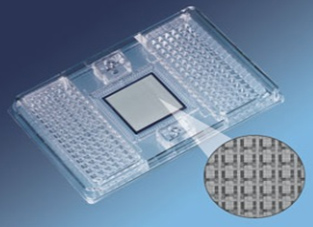
| The aim of this project is to characterize at molecular level markers defined in the endometrial receptivity molecular signature, taking into account factors necessary for endometrial receptivity and for immunologic response control; for the purpose of developing a new diagnostic tool for endometrial receptivity state. The chosen platform for this new diagnostic tool is BioMark 96.96 IFC Dynamic Array of Fluidigm. This new real time quantitative PCR (qRT-PCR) platform is based in TaqMan probes, it needs less reactive amount and less pippeting steps than other methods, it uses nanometric scale and, most important, it allows the simultaneous study of 96 genes in 96 samples. Thus, qRT-PCR analysis in this platform is robust, simpler and more affordable. |
Figure 2. Image of the platform 96.96 IFC Dynamic Array from Fluidigm. |
Induction of apoptosis on endometrial stromal cells under treatment with copperCopper intrauterine device (IUD) releases an amount of copper in the human endometrial tissue that induces physiological and biochemical changes in the endometrial cells. This method has side effects (mostly pain and bleeding). Copper, an essential element, is toxic when present in excess in mammalian tissues and could trigger apoptosis. During the execution phase of apoptosis, the apoptotic microtubule network (AMN) is formed to prevent cell content leakage into the extracellular space. | |
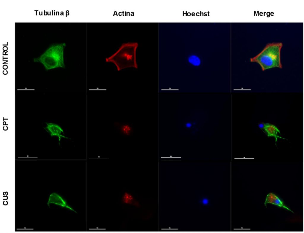
| The objective was to investigate the apoptotic effect of copper on human endometrial stromal cells at the same concentration than copper released by IUD and study the presence of the AMN. Thus, stromal cells obtained from endometrial biopsies (normal woman) were divided into 5 groups: control group (CNT), 10µM Camptothecin (positive control — apoptosis inducer, CPT) and the three different concentration of copper release reproducing the different phases of the menstrual cycle. Analysis was done by counting the percentage of apoptotic cells identified by immunofluorescence using three different markers: b-tubulin, actin and nucleus morphology. Apoptosis quantification was assessed by observing nuclei fragmentation, lack of actin cytoskeleton, and/or cell morphology by phase contrast microscopy. By this way, we showed that the three different concentrations of copper release during the menstruation had apoptotic effect. The apoptosis quantification showed that there are differences between the control group and the three copper groups (ANOVA P < 0.05). However, the level of apoptosis did not increase with time of exposure. Likewise, no significant differences were observed among copper groups. The amount of copper released by the IUD induces a higher percentage of apoptosis on these cells. These findings could explain the higher abnormal uterine bleeding in these subjects. |  Figure 2. AMN formation, actin cytoesqueleton absence and nuclear fragmentation (tagged with Hoechst) at 48h after copper treatment (CUS). CPT: camtotecine, citotoxic drug used to positive control. |
Expression analysis of tumor markers in human tumor samples to create metastasis free survival predictors and to know the detoxification cellular stateLast dates from WHO show an incidence of 14.1 millions of new cancer cases and 8.2 millions of cancer deaths. Dividing the population by sex, lung cancer presents the highest mortality rate in men and breast cancer, in women. Global gene expression analysis of tumor tissues has allowed us to identify characteristic expression patterns of a tumor type. Due to these transcriptomic imprintings, different prognostic markers have been developed helping in the generation of predictor tests that separate patients in different risk groups, predict tumor evolution or response to a treatment. Nowadays, several tests have been validated by FDA in USA, such as Ampliseq, Oncotype Dx and MammaPrint. However, it is needed to develop new more economic, simpler and more efficient tests that give us more information to design more efficient chemotherapeutic protocols. The aim of this project is to generate 3 predictor tests:
| |
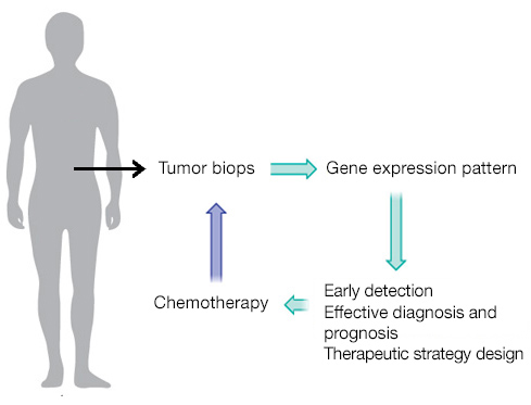
| The chosen platform for this new diagnostic tool is BioMark 96.96 IFC Dynamic Array of Fluidigm. This new real time quantitative PCR (qRT-PCR) platform is based in TaqMan probes, it needs less reactive amount and less pippeting steps than other methods, it uses nanometric scale and, most important, it allows the simultaneous study of 96 genes in 96 samples. Thus, qRT-PCR analysis in this platform is robust, simpler and more affordable. The probes designed to analyze gene expression of each target in a quantitative manner have been validated, and we have obtained cDNA from total RNA extracted from lung cancer biopsies. Thus, once all cDNA from breast cancer biopsies is available, we will carry out gene expression analysis of all markers and several statistic analyses will be done to set up the predictor tests. Figure 3. Genetic prognosis scheme in cancer (adapted from Lujambio et al, 2012). |  |
Ectopic pregnancy biomarkers in peripheral bloodEctopic pregnancy, a condition where the pregnancy occurs outside the endometrial cavity, is the first cause of maternal morbidity and mortality in the first trimester. The leading risk factors are anatomic damage in fallopian tubes, previous surgeries, infections and smoking. This risk is higher in assisted reproductive treatments and in vitro fecundation. However, a great amount of ectopic pregnancies occur in women without risk factors. The major complication in ectopic pregnancy is internal hemorrhage, which could lead to death. In USA, 1.3% of pregnancies are ectopic and last epidemiological dates estimate that 6% of death in pregnant women is due to ectopic pregnancies; these dates can be extrapolated to European population. | |
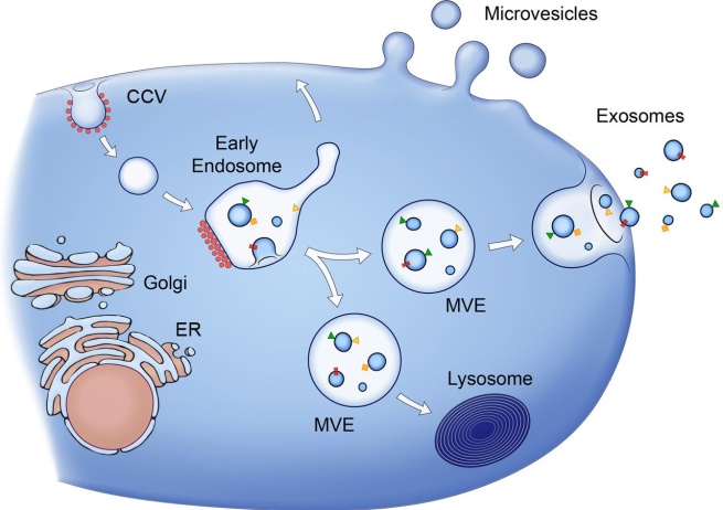
| Early diagnosis of ectopic pregnancy greatly improves morbidity prognosis. Currently, the only possible diagnosis is based on serum β-hCG and ultrasound findings. However, a follow-up for several days until reach a final diagnosis is common, generating an annual cost of 9 million pounds in the UK. Thus, many researchers are searching for a reliable and easy to use marker of ectopic pregnancy. Exosomes are nanovesicles secreted by cells after the fusion of internal vesicles with plasmatic membrane. The molecular content of exosomes represents the cellular type that has developed each exosome and their physiological status. Exosomes are in corporal fluids, especially in blood and urine; and they can be used as diagnostic tools in many types of cancer, neurodegenerative diseases and other pathologies. Figure 4. Exosomes and extracelular microvesicles (MVE) biogenesis (Raposo and Stoorvogel, JCB 2013). |  |
| This project aims to characterize markers from exosomes in peripheral blood of women with ectopic pregnancy using omics methods, and employing these markers for the early diagnosis of ectopic pregnancy. This could have a great impact in our society, diminishing pregnancy mortality, surgeries, lost of fertility, and, at the same time, decreasing the cost of currently diagnostic systems. Nowadays, whole genome gene expression microarrays have been performed with own funds (Whole Genome 44K, Agilent Technologies) from total RNA of the maternal blood of women with ectopic (n=6) and uterine pregnancies (n=6). This analysis revealed a list of 57 differential expressed genes, 44 up-regulated and 13 down-regulated with a fold change greater than 2.0 (p-value<0.05). Moreover, we have designed the probes for each target and validated the results obtained in microarrays through quantitative PCR of some genes: ORM 1, ORM 2 and UTS2, using the same samples in which the microarray analysis was done: ectopic pregnancy (n=6) and uterine pregnancy (n=6). | |
